What to Expect from Cataract Surgery

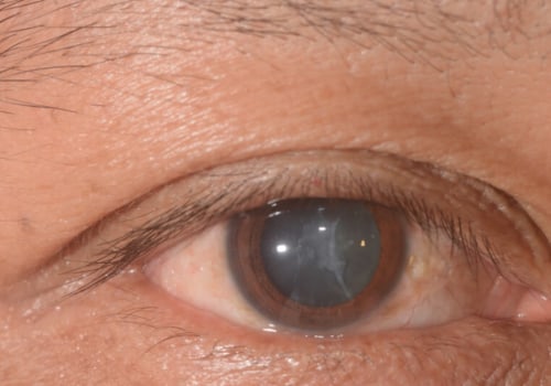
Cataract surgery is a common operation to remove the natural lens of the eye and replace it with an intraocular lens. It is a complex procedure that requires a great deal of care, and can be painful, but the benefits outweigh the risks. Knowing what to expect before, during, and after the surgery is essential for getting the best treatment possible.

Symptoms of Cataracts

The most common symptom of cataract surgery is blurry vision due to the clouding of the lens. This reduces the amount of light that reaches the retina, and may be accompanied by an increased need for glasses. During the procedure, an incision is made in the cornea and ultrasound energy is used to make an opening in the anterior portion of the lens. Immediately after, patients may experience slight bleeding, but if it continues or worsens, they should see a medical professional.

Recovery Time

It can take up to a few days for vision to return to normal due to swelling and inflammation. If eyesight is still blurry, try driving until it's settled. Other unusual symptoms include double vision, nausea, fever, and drowsiness. The pupil will remain dilated for several hours, causing close-up vision to be blurry. If there is no pain, patients can usually go home the same day.

Slit-Lamp Test

The slit-lamp test is used to detect fluid pressure in the eye and any other signs of damage. A protective shield may be placed over the eye if there is a moderate leak. Bandage contact lenses or cycloplegic inhibitors may also be used.

Procedure for Cataract Surgery

Cataract surgery is an outpatient procedure that removes a cloudy natural lens from your eye and replaces it with a clear artificial lens. It is a very effective and safe procedure, but can have complications if proper postoperative care isn't taken. After the procedure, patients must use the correct eye drops and medications to prevent infection. They should also wear an eye shield during sleeping and sunglasses outdoors. The first step of the procedure is to make a small incision on the edge of the cornea without stitches. An ultrasonic handpiece is then used to emulsify the eye's internal lens and break it into smaller pieces. A clear intraocular lens is then inserted behind the iris and set for near, intermediate, or distance vision according to patient needs. The eye is numbed with anesthetic drops and an eyelid holder prevents blinking during the 15-minute procedure. The ophthalmologist will then insert the lens into the eye capsule and close up the incision with stitches or glue.

Leave Message

Your email address will not be published. Required fields are marked *